This page collects published research, endoscopic images, and video showing Necator americanus (NA), the human hookworm, in the gastrointestinal tract.
Its primary purpose is visual: to provide examples of what adult NA can look like during endoscopy and make the original sources easy to find.
Adult Necator americanus are small worms that attach to the intestinal mucosa. Their appearance can vary considerably depending on their position, whether they are moving, whether they are attached or feeding, and the angle of the endoscope.
Published endoscopic images and video show worms that may appear curved, tightly curled, loosely coiled, or relatively extended. Against the intestinal mucosa they are generally pale and visually distinct.
The image below shows a mature male and female N. americanus attached to healthy duodenal mucosa with prominent villi, alongside earlier feeding and ulceration stages from the same study.

Additional video-frame stills of confirmed NA in the duodenum appear in the JACI section below.
Additional examples appear in the sources below.
Experimental hookworm infection and gluten microchallenge promote tolerance in celiac disease
Croese J, Giacomin P, Navarro S, et al.
Journal of Allergy and Clinical Immunology. 2015;135(2):508–516.
DOI: 10.1016/j.jaci.2014.07.022
Full text: jacionline.org/article/S0091-6749(14)01010-0/fulltext
This 52-week study followed 12 adults with diet-managed celiac disease who were deliberately inoculated with 20 Necator americanus larvae and subsequently exposed to escalating amounts of gluten.
All participants developed established NA infections. The researchers confirmed infection by detecting hookworm eggs in feces throughout the study.
Of particular relevance here, the researchers also directly observed mature Necator americanus during endoscopy. Adult worms, sometimes mating, were visualized in the duodenum.
The article includes supplementary endoscopic video showing the living worms in the duodenum.
Endoscopic video
Video E1: Mature Necator americanus in the duodenum
Selected frames:
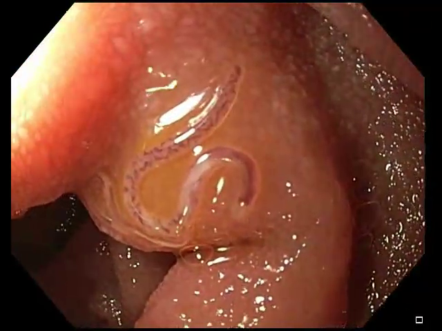
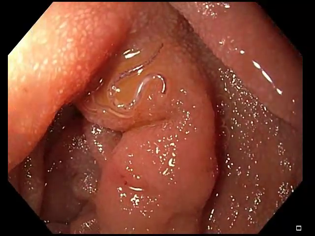
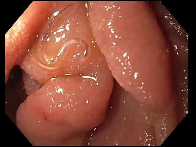
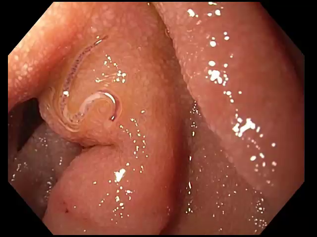
The video is particularly useful as a visual reference because movement and changing orientation make it easier to distinguish the worms from intestinal folds, mucus, or other structures.
Study findings
The study was designed primarily to investigate whether controlled NA infection could modify the immune response to gluten in people with celiac disease.
Participants underwent progressively larger gluten challenges while researchers monitored symptoms, serology, intestinal histology, and immune responses.
The authors reported that NA infection combined with gradual gluten microchallenge was associated with increased gluten tolerance and stabilization or improvement in the measured indices of gluten toxicity. Intestinal inflammatory T-cell responses changed following hookworm infection, including a reduction in IFN-γ-expressing intestinal T cells and an increase in regulatory T cells.
For the purposes of this page, the study is particularly valuable because the identity of the organism is known: participants were deliberately inoculated with Necator americanus, established infections were independently confirmed, and mature worms were subsequently visualized in the duodenum during endoscopy.
Changed gluten immunity in celiac disease by Necator americanus provides new insights into autoimmunity
Croese J, Gaze ST, Loukas A.
International Journal for Parasitology. 2013;43(3–4):275–282.
DOI: 10.1016/j.ijpara.2012.12.005
Full text: sciencedirect.com/science/article/pii/S0020751912003219
Graphical abstract: ars.els-cdn.com/…/fx1_lrg.jpg
This review describes experimental human infection with Necator americanus and summarizes clinical research examining its effects on immune responses in people with celiac disease.
The authors describe NA as a durable human gastrointestinal nematode and discuss their experience deliberately establishing controlled infections in research participants.
The paper reports that experimental NA infection can be reliably established in a clinical setting and describes low-dose infection as generally well tolerated in the studies reviewed. It also examines how the presence of NA alters mucosal immune responses, including responses to gluten.
Endoscopic appearance
The paper contains particularly useful visual documentation of adult hookworms in the intestine.
Figure 5 includes images of feeding hookworms associated with healthy-appearing duodenal mucosa.

These images are useful because they show the worms in the anatomical environment in which an upper endoscopist might encounter them. The wider field also provides a useful impression of their scale relative to the intestinal lumen and mucosal folds.
Helminthic Therapy Wiki: ulcerative colitis case history
The effects of NA and TTO on ulcerative colitis
This page documents the experience of an individual with longstanding ulcerative colitis who used both Necator americanus (NA) and Trichuris trichiura ova (TTO).
The individual reported beginning helminthic therapy after 11 years with ulcerative colitis and eventually experiencing sustained remission. The page follows the case over a number of years and includes later colonoscopy findings.
The account reports continued use of both NA and TTO and includes colonoscopy images taken after years of remission. The accompanying medical findings describe normal mucosa throughout the colon and normal terminal ileum, with biopsies described as unremarkable and without evidence of active or chronic ileitis or colitis.
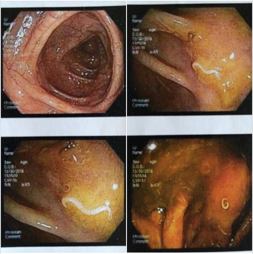
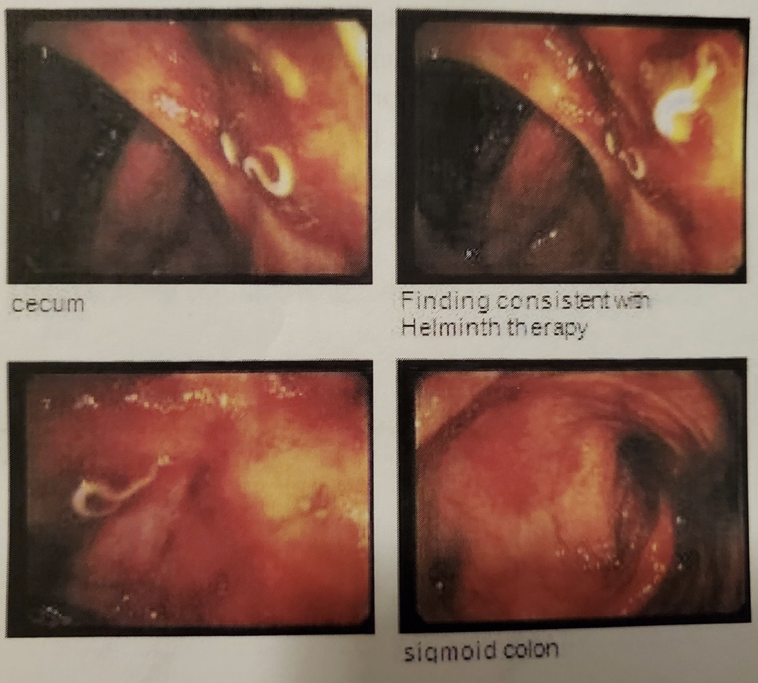
Important identification note
The source for this image — a patient’s own Facebook post — identifies these worms as whipworms, but that identification wasn’t clinically confirmed. Species can’t be reliably determined from a single endoscopic still, since NA and Trichuris can look similar depending on which segment of the body is visible. This image is included for general GI visual context; it shouldn’t be treated as confirmed NA identification either.
Helminthic Therapy Wiki: Crohn’s colitis case history
The effects of NA and TTO on Crohns colitis
This page documents a second individual who had severe Crohn’s colitis until 2014, then began helminthic therapy using both NA and TTO and reached remission within 3–6 months. He returned to his GI for a colonoscopy in early 2019, roughly 5 years after starting therapy, and reported that his doctor described seeing no signs of active disease beyond scarring from old inflammation, along with a casual mention of seeing worms present.
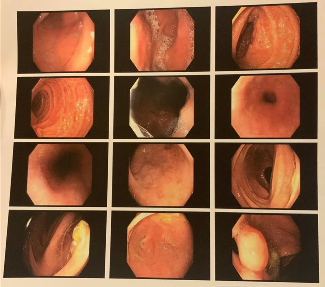
Post-remission colonoscopy imagery; no clearly identifiable worm is visible in the images themselves. Included for general context on long-term mucosal appearance under combined NA/TTO therapy, not as a species identification reference. As with the UC case above, the patient’s mention of seeing worms during the procedure is a casual paraphrase of his GI’s comment, not a clinical species determination — and since he was dosing both NA and TTO, it can’t be attributed to either species specifically.
Additional endoscopic images
The following images have been collected as additional visual examples.
They are retained here because different photographs emphasize different features: overall size, curvature, attachment, orientation, and appearance against the intestinal mucosa.
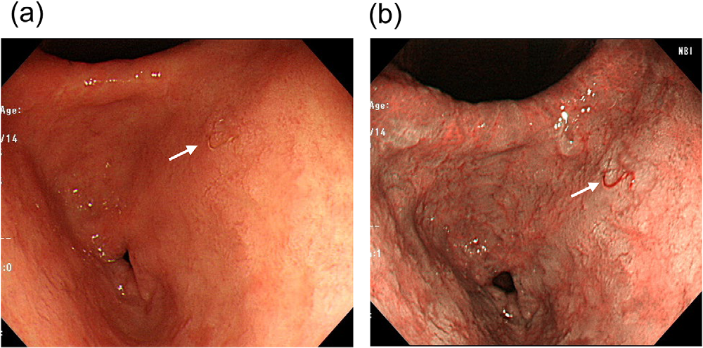
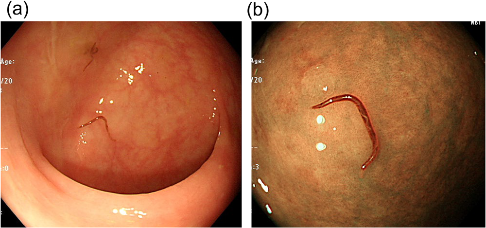
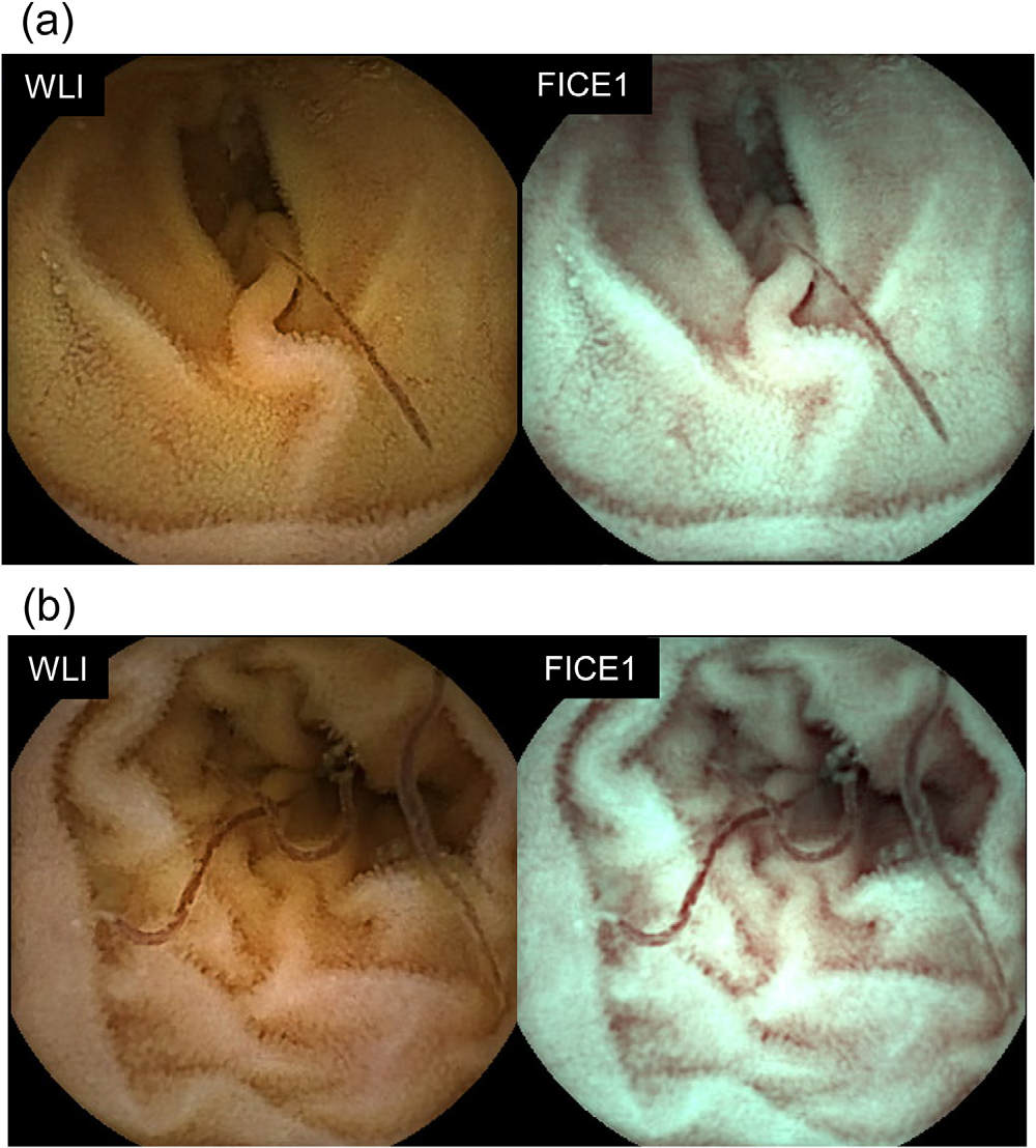
Where possible, each image should retain its original source information so that its provenance can be verified.
Distinguishing the sources
The strongest visual evidence for identifying Necator americanus comes from the controlled human-infection research.
In those studies:
- the organism administered was specifically Necator americanus;
- participants received known numbers of NA larvae;
- established infection was confirmed independently;
- mature worms were directly observed during endoscopy; and
- published images and supplementary video document their appearance in the duodenum.
The JACI supplementary video is particularly useful because a moving worm can be recognized in ways that are sometimes less obvious in a single still photograph.
Sources
Croese J, Giacomin P, Navarro S, et al. Experimental hookworm infection and gluten microchallenge promote tolerance in celiac disease.
Journal of Allergy and Clinical Immunology. 2015;135(2):508–516.
jacionline.org/article/S0091-6749(14)01010-0/fulltext
DOI: 10.1016/j.jaci.2014.07.022
Croese J, Gaze ST, Loukas A. Changed gluten immunity in celiac disease by Necator americanus provides new insights into autoimmunity.
International Journal for Parasitology. 2013;43(3–4):275–282.
sciencedirect.com/science/article/pii/S0020751912003219
DOI: 10.1016/j.ijpara.2012.12.005
Helminthic Therapy Wiki. The effects of NA and TTO on ulcerative colitis.
helminthictherapywiki.org/wiki/The_effects_of_NA_and_TTO_on_ulcerative_colitis
Helminthic Therapy Wiki. The effects of NA and TTO on Crohns colitis.
helminthictherapywiki.org/wiki/The_effects_of_NA_and_TTO_on_Crohns_colitis
Croese J, Wood MJ, Melrose W, Speare R. Allergy Controls the Population Density of Necator americanus in the Small Intestine.
Gastroenterology. 2006;131(2):402–409.
DOI: 10.1053/j.gastro.2006.05.019
Takebe T, Osawa S, Watanabe Y, Yamagata S, Aoyama T, Kawamura Y, Manabe A, Kisamori T, Sano M, Sugimoto K. Endoscopic Diagnosis of Necator americanus Infection Presenting With Persistent Iron-Deficiency Anemia: Usefulness of Image-Enhanced Endoscopy and Capsule Endoscopy.
DEN Open. 2027;7:e70352.
DOI: 10.1002/deo2.70352